The knee
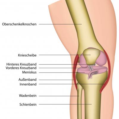
The knee joint is the largest joint of the human body. The thighbone (femur), the shin (tibia) and the kneecap (patellar) form the bony joint partners.
The knee joint is a composed joint. It consists of two single joints, the kneecap joint, which is located between thighbone and kneecap, and the popliteal fossa joint, which is located between thighbone and tibial plateau.
At the back of the knee joint the hollow of the knee is located, in whose depths important blood vessels and nerves run through.
The knee joint is the actual joint responsible for the diffraction of the knee. It is a mixture of a wheel and hinge joint. One labels it as a rotation hinge or bi-condylar joint. It makes the bending and stretching possible as well as a slight in- and outward twist of the knee bent at an angle of 90 degrees. It is located between the thighbone curved outward and the shin plateau. It has to withstand great stresses, but make a sufficient agility possible at the same time.
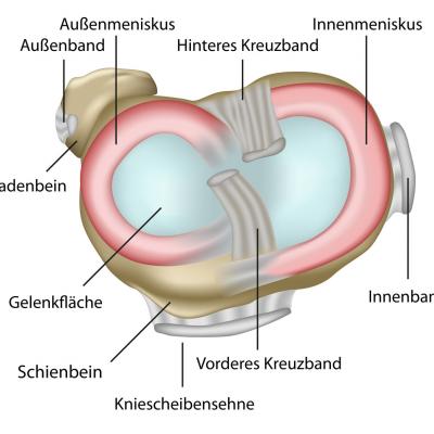
As the joint surfaces (articular) connected with each other do not fit perfectly together, this inequality (incongruity) is equalized by crescent-shaped fibrous cartilage disks, the menisci, which can follow the swivel movements. Another task of the menisci is to enlarge the contact surface between shin and thighbone.
One distinguishes between an inner meniscus, which is c-shaped, which is larger and a little bit less movable (because it is grown together with the inner ligament), and an outer meniscus, which has the form of a circle, which is smaller and more movable (because it is not grown together with a sideband). In the cross section the menisci are v-shaped. The high edge lies on the outside, the low one inside. As the thighbones lie exactly in the middle of the shin plateau and peripherally on the menisci, they bear an essential part of the load.
Moving the knee joint, the menisci are pushed ahead by the thighbones. During bending, the bones roll back and press the menisci backwards, during stretching, they come back forward. During an outward rotation of the lower leg the outside meniscus is pushed over the shin to the front, the inside meniscus pulled back. In case of an inward rotation it is the other way round.
At each of their posterior and anterior horns the menisci are connected with the plateau of the shin via a short and strong retaining ligament. In addition to these four horns at the shin, the menisci are connected with the inner thighbone via variably applied ligaments.
The bone partners are in a very close contact to each other. In order that a painless and undisturbed flexibility of the knee joint is also possible on the contact surfaces, they are (like all joint surfaces in the human body) covered with a very smooth and whitish cartilage layer, the so-called hyaline cartilage.
The central and most important stabilization of the knee joint to the front and backwards is undertaken by the cruciate ligaments. The lateral stabilization is provided by the internal and external sideband. The shock-absorber function inside of the knee joint is undertaken by the inner and outer meniscus, which also function as secondary stabilizers.
The anterior cruciate ligament, the inner ligament as well as the inner and outer meniscus, which stabilize the knee joint together, are particularly often affected by injuries.


