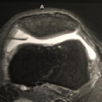
The Plica syndrome
Like any other joint, the knee joint is coated with a thin and smooth mucosa (synovia). The synovia produces joint fluid, which reduces the friction within the joint and supplies the joint cartilage with nutrients. During the embryonic phase the synovia develops a membrane, which divides the knee joint into two seperate areas.
Usually, the membrane recedes completely until the end of puberty for the benefit of a greater mobility inside of the knee joint.
Among approx. 50-70% of the adults, a small or also greater fold, a so-called plica, remains. Most of the time it is located behind, above or inside (=medial) of the kneecap.
Many people with a plica have no problems at all. If the plica is embossed though, irritations can appear. Particularly an overstress of the knee joint leads to an irritation of the plica and thus to the so-called plica syndrome. The most frequent reasons are stressful activities during which the knee is repeatedly bent and stretched again (like running, cycling or exercises on so-called steppers) or fabric loads (hit, fall) as well.
The plica itself, but also the tissue surrounding it, swells or bleeds in and becomes painful. This thickening then rubbs on the cartilage inside of the knee joint and can – in case of continuous load – lead to an inflammation of the joint (=arthritis) and later on to a cartilage wear off.
The most frequent complaints in case of the plica syndrome are:
- pains during load, mostly on the in- or backside of the kneecap
- creaking or crackling of the joint in a specific position while bending the joint
- a feeling of a blocking during the stretch movement
- stiffness of the joint after prolonged sitting
- in some cases the thickened plica is also touchable under the skin, or it comes to a swelling of the whole knee joint


